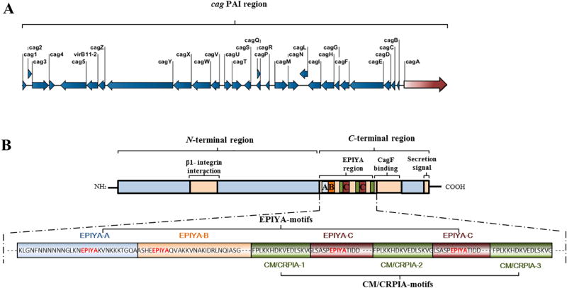
Figure 1. Overall structure of the cag PAI (A) and CagA protein (B) in H. pylori strain P12.
A. Structure of the cag PAI region (~37 kb). The region comprises 28 genes that encode components of the cag-T4SS, including the CagA protein effector.
B. Structure of the CagA protein (1,214 amino acid residues). The N-terminal part of CagA harbors a putative β-integrin-binding region. The C-terminal region comprises the EPIYA region, which contains EPIYA ABCC motifs and three MKI/CM/CRPIA motifs, regions that bind to the secretion chaperone CagF and contain the C-terminal secretion signal.

