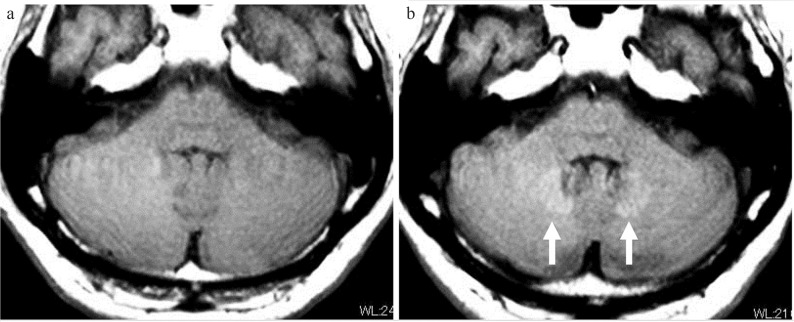
Fig. 1.
Deposition of gadolinium in the dentate nucleus. A case with multiple sclerosis between an initial MRI performed in 2009 (a) and an MRI performed in 2017 (b), repeated contrast-enhanced MRI examinations were performed 17 times using linear-type gadolinium-based contrast medium (GBCM) five times and macrocyclic GBCM 12 times. On the T1-weighted image obtained with fast spin echo imaging in 2017 (b: arrow), the bilateral dentate gyrus shows high-signal intensity. The signal intensity ratio between the pons and dentate nucleus was 1.02 in 2009 and 1.08 in 2017.

