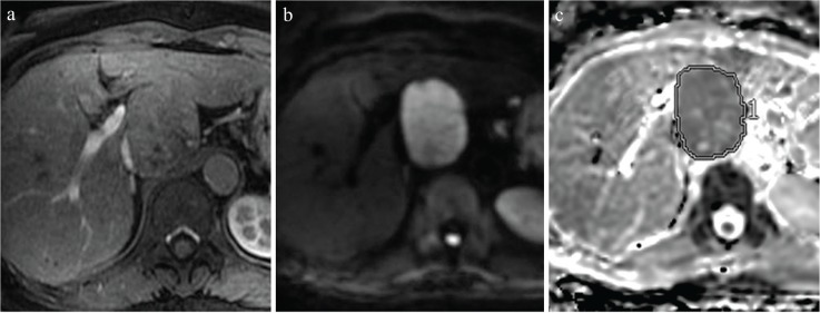
Fig. 2.
Rules for ROI placement, as demonstrated on representative images from a 69-year-old woman with hepatitis B. (a) Hepatic arterial-phase images show a slightly enhanced tumor at the caudate lobe. (b) This nodule shows high intensity on a diffusion-weighted image (b = 1000 s/mm). (c) We manually placed ROIs on the entire lesion on apparent diffusion coefficient (ADC) maps using SYNAPSE VINCENT software (FUJIFILM Medical, Tokyo, Japan).

