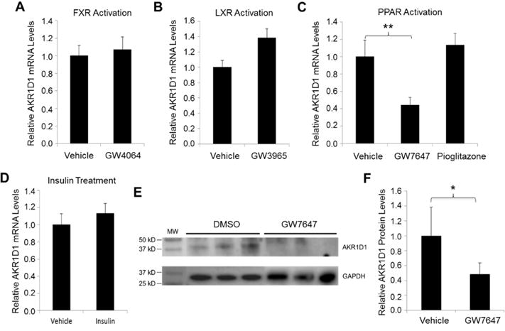
Fig. 9. Activation of PPARα signaling repressed AKR1D1 expression in vitro in HepG2 cells.

(A) HepG2 cells were treated with synthetic FXR agonist GW4064 (1μM), (B) LXR agonist GW3965 (1μM), (C) PPARα agonist GW7647 (10μM) or PPARγ agonist pioglitazone (10μM) or (D) insulin (100ng/ml) for 30h, followed by detection of AKR1D1 mRNA expression by TaqMan real-time PCR. The expression levels of GAPDH were used to normalize the expression levels of AKR1D1 for the treatments with GW4064, GW3965, GW7647 and pioglitazone. The expression levels of β-actin were used to normalize the expression levels of AKR1D1 for the treatment with insulin. (E) HepG2 cells were treated with PPARa agonist GW7647 for 48h, followed by detection of AKR1D1 protein. A representative Western blot showing protein expression of AKR1D1 and GAPDH. (F) quantification of AKR1D1 protein levels on Western blots normalized by the levels of GAPDH protein. For both real-time PCR assays and Western blot, the experiments were independently repeated at least once with each experiments having triplicates for each treatment. ** p<0.01 with one-way ANOVA followed by Tukey post-hoc test. * p<0.05 with Student’s t-test.
