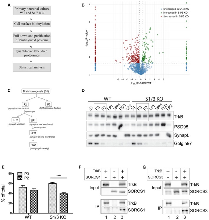
Figure 7. Altered subcellular localization of TrkB in primary neurons and brains from S1/3 KO mice.

-
AWorkflow of the cell surface proteome analysis in primary neurons.
-
BResults of quantitative label‐free proteomics comparing the surface proteomes of WT and S1/3 KO primary cortical neurons. Plot represents –log10(P‐value) and log2 (relative levels S1/3KO/WT) obtained for each protein. For proteins that were detected in either S1/3 KO or WT samples only, the log2(S1/3 KO/WT) was set to 10 or ‐10, respectively. Threshold values are 1.3 for –log10(P‐value) and ±0.3 for log2(S1/3 KO/WT). Proteins showing higher (green) or lower (red) surface levels in S1/3 KO as compared to WT neurons are indicated. Proteins with unchanged cell surface levels are indicated in blue. n = 3 biological replicates/group (with each biological replicate run in two technical replicates).
-
CScheme of brain membrane fractionation used to analyze subcellular localization of TrkB in vivo (see method section for details).
-
DRepresentative Western blotting of TrkB in various brain membrane fractions obtained as described in panel (C). Highlighted lanes represent P3 and P2 fractions, quantified in (E). Detection of PSD95, synaptophysin, and Golgin 97 was used to assess the accuracy of subcellular fractionation.
-
EQuantification of total TrkB distribution between P3 (light membrane fraction) and P2 (synaptosomal fraction) from Western blots exemplified in panel (D) (n = 5 mice/group, 7–12 weeks of age). The TrkB pool is shifted toward the light membrane (P3) fraction in S1/3 KO brains. Data are shown as mean ± SEM and analyzed using two‐way ANOVA with Bonferroni post‐test. ****P < 0.0001.
-
F, GCo‐immunoprecipitation of SORCS1 and SORCS3 with TrkB. TrkB was immunoprecipitated from transiently transfected Chinese hamster ovary cells and the presence of SORCS1 or SORCS3 in the immunoprecipitates documented by Western blotting. Panel input shows levels of the indicated proteins in the cell lysates prior to immunoprecipitation. Panel IP documents co‐immunoprecipitation of SORCS1 (F) and SORCS3 (G) from cells expressing (lanes 3) but not from cells lacking TrkB (lanes 2). The experiment was replicated twice.
Source data are available online for this figure.
