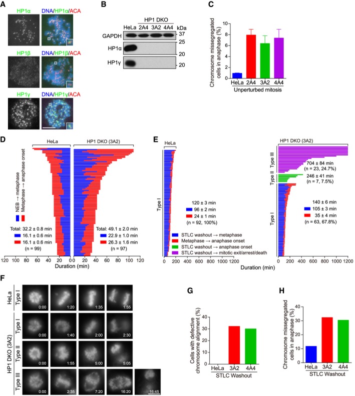
Figure 1. Double knockout (DKO) of HP1α and HP1γ causes defective mitosis progression.

-
AHeLa cells were treated with nocodazole for 3 h. Mitotic chromosome spreads were immunostained.
-
BAsynchronous HeLa and the indicated HP1 DKO clones were immunoblotted.
-
CHeLa and HP1 DKO clones were fixed and stained with anti‐human centromere autoantibodies (ACA) and DAPI. The percentage of cells with lagging chromosomes was determined in 200 anaphase cells. Means and ranges are shown (n = 2).
-
DThe mitosis progression of HeLa and HP1 DKO clone 3A2 cells stably expressing H2B‐GFP was analyzed by time‐lapse live imaging. The time from nuclear envelope breakdown (NEB) to metaphase chromosome alignment, and from metaphase to anaphase onset, was determined. Fate profiles of cells were determined. See also Movies EV1 and EV2.
-
E, FHeLa and HP1 DKO clone 3A2 cells stably expressing H2B‐GFP were released from 5‐h treatment with STLC, followed by live imaging of mitosis progression. Fate profiles of mitotic cells (E), and the selected frames of the movies (F), are shown. Time stated in hours:minutes. See also Movies EV3,EV4 and EV5.
-
G, HThe percentage of mitotic cells with defective chromosome alignment (G) and chromosome missegregation (H) were quantified in 102 cells (for HeLa and clone 3A2) and 74 cells (for clone 4A4).
