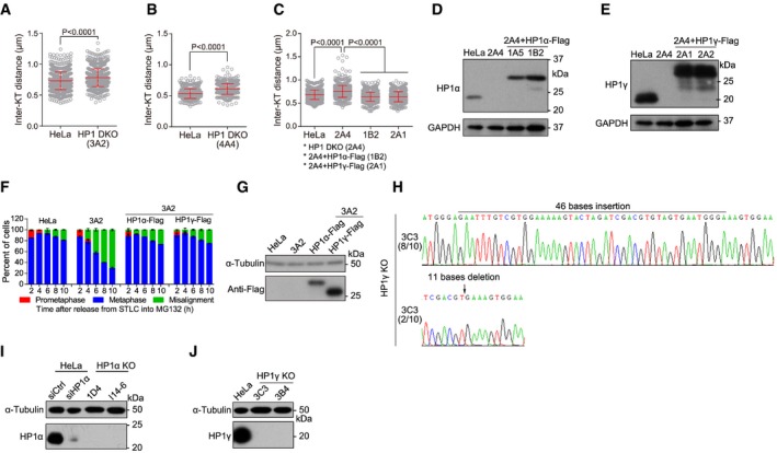
Figure EV2. HP1α and HP1γ are redundantly required to protect mitotic centromere cohesion (related to Fig 2).

-
A–CThe indicated stable cell lines were treated with nocodazole for 3 h. Mitotic chromosome spreads were stained with CENP‐C or ACA antibodies and DAPI. The inter‐KT distance was measured on over 400 chromosomes in over 20 cells (unpaired t‐test). Means and SDs are shown.
-
D, ELysates of asynchronous HeLa and HP1 DKO clone 2A4 cells with or without stable expression of the indicated exogenous HP1 were immunoblotted.
-
F, GHeLa and HP1 DKO clone 3A2 cells with or without transient expression of HP1α‐Flag or HP1γ‐Flag were analyzed for metaphase chromosome alignment in around 200 cells. Means and ranges are shown (F; n = 2). Lysates of asynchronous cells were immunoblotted (G).
-
HGenomic DNA sequencing of HP1γ KO clone 3C3 cells. The genomic DNA PCR fragments were subcloned and sequenced. Eight out of 10 bacterial colonies showed insertion of 46 bases, whereas the rest two bacterial colonies showed deletion of 11 bases.
-
I, JLysates of asynchronous HeLa and HP1α or HP1γ KO cells were immunoblotted.
Source data are available online for this figure.
