-
A
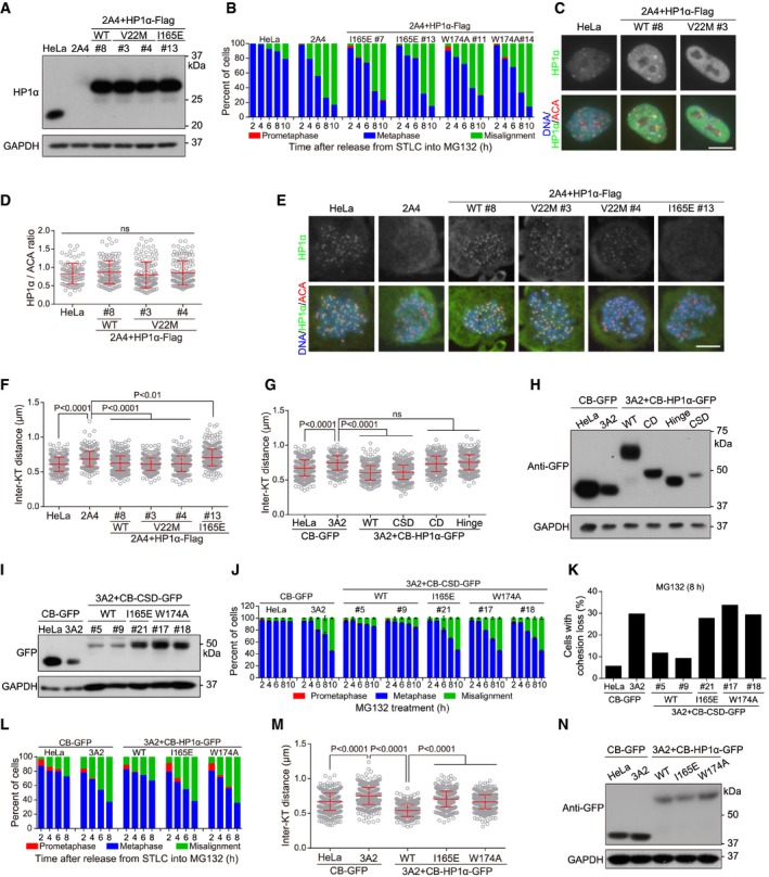
Lysates of asynchronous HeLa and HP1 DKO clone 2A4 cells with or without stable expression of the indicated exogenous HP1α were immunoblotted.
-
B
HeLa and the indicated stable cell lines were released from 5‐h treatment with STLC into MG132‐containing medium, fixed at the indicated time points for DNA staining, and then quantified in around 100 cells.
-
C
Asynchronous HeLa cells and the indicated stable cell lines were immunostained.
-
D, E
HeLa and the indicated stable cell lines were treated with nocodazole for 3 h. Mitotic chromosome spreads were immunostained. The centromeric HP1α/ACA immunofluorescence intensity ratio was determined on over 100 chromosomes in 10 cells (D) (unpaired t‐test). Example images are shown (E).
-
F
HeLa and the indicated stable cell lines were treated with nocodazole for 3 h. Mitotic chromosome spreads were stained with ACA antibodies and DAPI. The inter‐KT distance was measured on over 400 chromosomes in over 20 cells (unpaired t‐test).
-
G
HeLa and HP1 DKO clone 3A2 cells transiently expressing the indicated fusion proteins were treated with nocodazole for 3 h. Mitotic chromosome spreads were stained with CENP‐C antibodies and DAPI. The inter‐KT distance was measured on over 400 chromosomes in over 20 cells (unpaired t‐test).
-
H
Lysates of asynchronous HeLa and HP1 DKO clone 3A2 cells transiently expressing the indicated fusion proteins were immunoblotted.
-
I
Lysates of asynchronous HeLa and HP1 DKO clone 3A2 cells stably expressing the indicated fusion proteins were immunoblotted.
-
J
The indicated stable cell lines were analyzed for metaphase chromosome alignment in around 200 cells. Means and ranges are shown (n = 2).
-
K
The indicated stable cell lines were analyzed for cohesion loss in around 100 cells.
-
L
HeLa and HP1 DKO clone 3A2 cells transiently expressing the indicated fusion proteins were released from 5‐h treatment with STLC into MG132‐containing medium, fixed at the indicated time points for DNA staining, and then quantified in around 100 cells.
-
M
HeLa and HP1 DKO clone 3A2 cells transiently expressing the indicated fusion proteins were treated with nocodazole for 3 h. Mitotic chromosome spreads were stained with CENP‐C antibodies and DAPI. The inter‐KT distance was measured on over 400 chromosomes in over 20 cells (unpaired t‐test).
-
N
Lysates of asynchronous HeLa and HP1 DKO clone 3A2 cells transiently expressing the indicated fusion proteins were immunoblotted.
Data information: Means and SDs are shown (D, F, G, J, and M). Scale bars, 10 μm.

