Fig. 1.
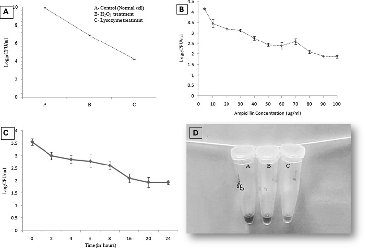
Characteristic features of Persister cells. a Exponential phase S. Typhi cells were subjected to environmental stress (10 mM of H2O2 and 22.5 μg/ml of lysozyme, 15 min each) and enumerated by plating on nutrient agar plates. b After environmental stress cells were further treated for 4 h with different concentrations (at MIC and higher than the MIC) of ampicillin. After 80 µg/ml no significant reduction in viability was recorded and these survived cells were taken as persister cells. c Time-dependent antibiotic (80 µg/ml) killing kinetics of S. Typhi cells showing biphasic curve; a characteristic property of persister cells. The initial reduction in the log10cfu/ml corresponds to sensitive cells followed by a plateau and fraction of cells that survived, indicating S. Typhi persister cells. d Metabolic state difference of persister cells under stress and normal condition: A—Persisters in PBS (Nutrient stress), B—Persisters in antibiotic stress (80ug/ml) and C—Persisters in nutrient media (no stress). Tube A and B do not show the change in color in comparison to tube C, where color is turned pink due to the reduction of resazurin (a component of dye) into red/pink color compound resorufin by metabolically active cells (color figure online)

