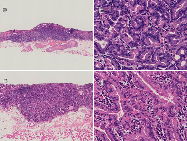
Figure 3.
A histological examination of the resected specimens (Hematoxylin and Eosin staining). Low magnification of the first lesion shows that its main portion is located in the deep layer of the lamina propria and that it has slightly infiltrated into the submucosa (a), whereas high magnification reveals that both lesions consist of fundic chief cell-like basophilic columnar cells (b and d). Low magnification of the second lesion shows that, although its main portion is located in the deep layer of the lamina propria, the entire lesion is confined within the mucosa (c).

