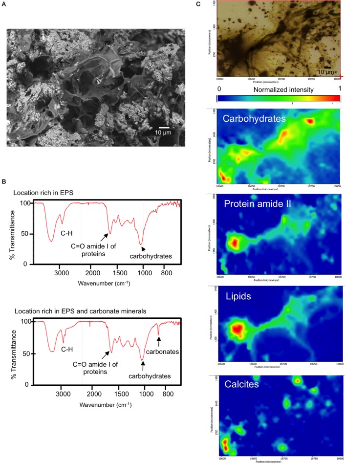
Figure 8.
(A) BAC (Bacalar microbialite) mineral inclusions and embedding organic matter (top, scanning electron micrograph), (B) SR-FTIR spectra of surface locations rich in EPS and EPS plus minerals of fresh BAC microbialite, (C) SR-FTIR spectromicroscopy images (200 μm by 150 μm) showing the distribution of microbes and minerals in a living BAC microbialite. Distribution heat maps of the protein amide II vibration modes at ~1,542 cm−1, the carbohydrates vibration modes at ~1,000 cm−1, calcite at ~870 cm−1, and lipid is base on the CH vibration modes near 2,900 cm−1. Scale bars: 10 μm. Transmittance is given in % units.

