
Fig 3.
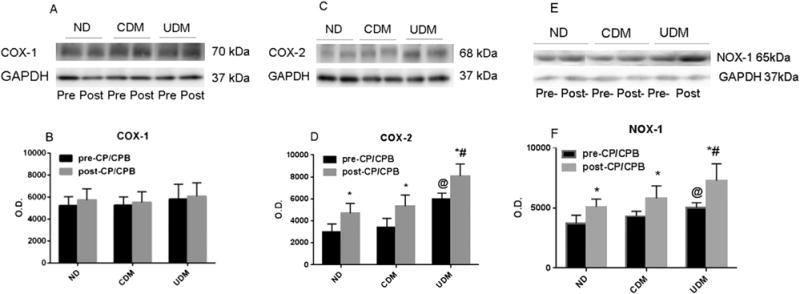
(A, C, E) Representative immunoblots of human atrial tissue samples from no diabetes mellitus (ND) patients, controlled diabetes (CDM) patients, and uncontrolled diabetes (UDM) patients. Lanes 1 to 6 loaded with 40 μg protein were developed for cyclooxygenase (COX)-1, COX-2, and nicotinamide adenine dinucleotide phosphate oxidase (NOX)-1 polypeptides. (GAPDH = glyceraldehyde 3-phosphate dehydrogenase.) (B) Densitometric evaluation of immunoblot band intensity shows no significant differences in the levels of COX-1 among three groups, or between pre-cardioplegia arrest and cardiopulmonary bypass (pre-CP/CPB [black bars]) and post-CP/CPB (gray bars). (D) Densitometric evaluation of immunoblot band intensity shows significant differences in the levels of COX-2 between UDM and ND or CDM groups, and between pre- and post-CP/CPB. (F) Densitometric evaluation of immunoblot band intensity shows significant differences in the levels of NOX-1 between UDM and ND or CDM groups, or between before and after CP/CPB. Data are mean ± standard error of the mean; n = 6 per group. *p < 0.05 versus pre-CP/CPB. @p < 0.05 versus ND pre-CP/CPB or CDM pre-CP/CPB. #p < 0.05 versus ND post-CP/CPB or CDM= post-CP/CPB. (O.D. = optical density.)