Figure 7.

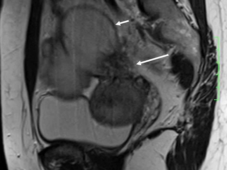
A 37-year-old woman with DIE. Sagittal T2-weighted image shows an irregular nodule (long arrow) with stellate margins in the right uterosacral ligament and an endometriotic cyst (short arrow).

A 37-year-old woman with DIE. Sagittal T2-weighted image shows an irregular nodule (long arrow) with stellate margins in the right uterosacral ligament and an endometriotic cyst (short arrow).