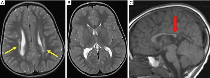
Figure 1.
Typical features of PVL with white matter scarring in the deep and periventricular white matter bilaterally (yellow arrows), reduced thalamic volume (middle picture) and reduction in the bulk of the cerebral white matter posteriorly (red arrow pointing at the reduced bulk of the corpus callosum posteriorly). This is a good candidate for SDR given the lack of extensive anterior white matter involvement and sparing of the basal ganglia structures. PVL, periventricular leukomalacia; SDR, selective dorsal rhizotomy.

