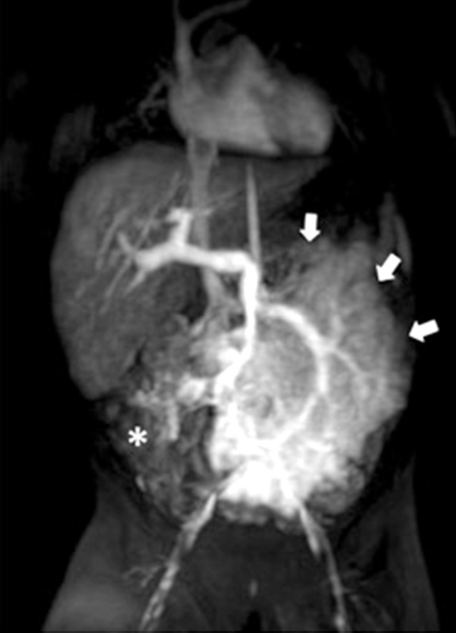
Figure 5.

Contrast-enhanced MRI, MIP coronal reconstruction, shows a “pseudo-nodular” tissue with inhomogeneous enhancement (arrows), which replaces omental fat and causes displacement of bowel loops to the peripheral side of the abdomen (asterisk). MRI, magnetic resonance imaging; MIP, maximum intensity projection.
