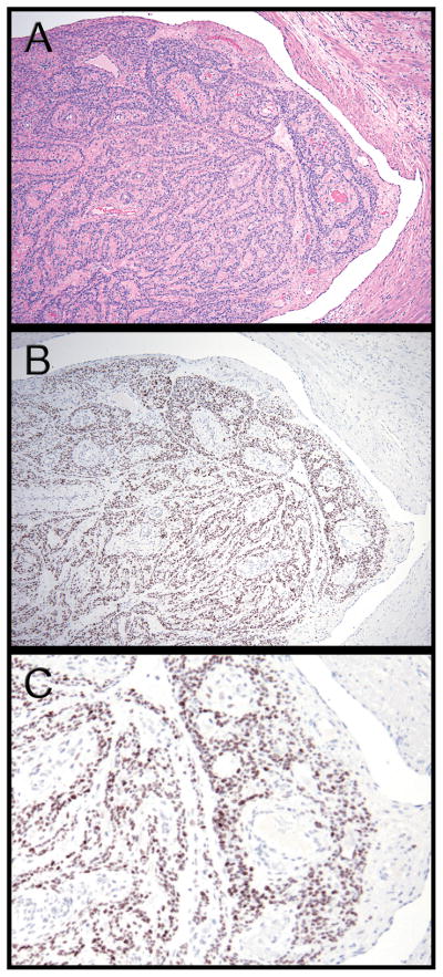
Fig. 1.
(A) Hematoxylin and eosin stained tissue section of Case 1 shows a plug-like tumor mass nearly occluding the residual cleft-like endothelial-lined vascular lumen with normal myometrium on the right. (B) Immunohistochemistry with a polyclonal HMGA2 antibody showing strong staining in intravenous leiomyomatosis tissue, but not in the adjacent myometrium. (C) Higher magnification image of panel B, in which one can appreciate that the HMGA2 staining is specific to nuclei in smooth muscle cells in lesional cells, but not in endothelial cells or smooth cells in the supporting normal blood vessels and adjacent myometrium.

