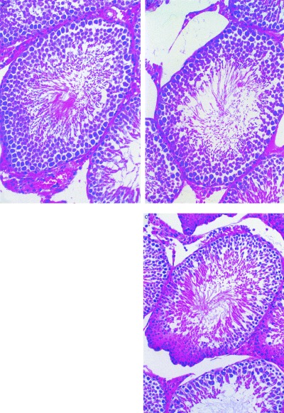
Figure 1.

Histological findings of the testes (hematoxylin & eosin stain). Control week 3 (a; reduced from ×66), experimental group week 3 (b; reduced from ×50), experimental group week 5 (c; reduced from ×80). The number of spermatogonia decreased in (b; 37) and (c; 27) compared with (a; 49). The number of pachytene spermatocytes decreased in (c; 30) compared to (a; 56) and (b; 58). Meanwhile, the number of Sertoli cells did not change in all specimens (approximately 35).
