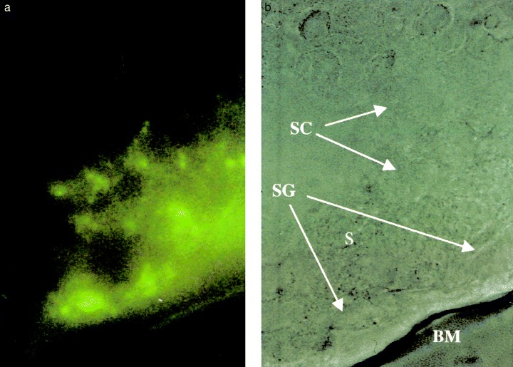
Figure 2.

(a) Oxine‐fluorescent cytochemistry of seminiferous tubules from rats in the experimental group at week 5 (left, reduced from ×1000). The same view without fluorescent light (right; BM, basement membrane; S, Sertoli cell; SC, spermatocyte; SG, spermatogonium). Cadmium was identified mainly at the cytoplasm of spermatogonia, and weakly at spermatocytes.
