
Fig. 2.
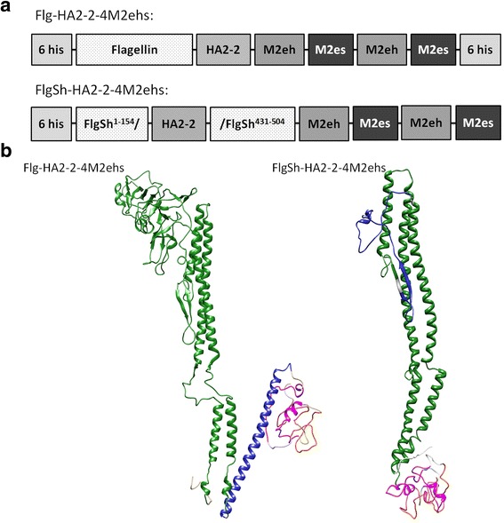
Structure (a) and theoretical modelling of 3D-structures (b) of monomeric recombinant fusion proteins Flg-HA2–2-4M2ehs and FlgSh-HA2–2-4M2ehs. Sizes of boxes are not drawn to scale. Yellow marks M2es; pink marks M2eh; blue marks HA2 fragment; and green is flagellin. Modelling performed with Phyre2 server, visualization made with USCF Chimera