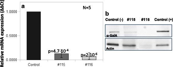
Fig. 1.

GLA mRNA levels (a) and Western blot of α-Gal A and actin (b) measured in leukocyte extracts of samples from subjects #115 and #116. In panel A, bars represent relative levels of mRNA expression (ΔΔCts) normalized to the control. Two different controls (healthy volunteers’ samples) were used in independent experiments (control bar). In panel B, Control (−) represents the sample from a healthy volunteer and Control (+) represents the sample from a patient diagnosed with FD carrying a missense pathogenic mutation
