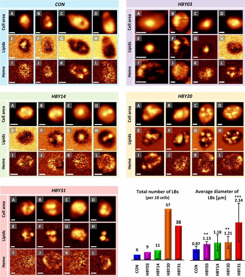
Fig. 5.

Distribution of selected components and LBs in yeast cells obtained via confocal Raman spectroscopy. In all panels: A–D the cell area is visualised by integration of the range 3050–2800 cm−1 (‘Cell area’); E–H LBs are shown by integral intensity of the band at 1444 cm−1 (‘Lipids’), I–J heme presence is shown by integral intensity of the band at 753 cm−1. Results of analysis of the presence of selected components are shown for single cells of all yeast cell lines: CAV (blue), HBY03 (purple), HBY14 (green), HBY20 (orange) and HBY31 (red). Each panel contains results obtained for 4 cells [in each panel: (A, E, I) cell 1, (B, F, J) cell 2, (C, G, K) cell 3, (D, H, L) cell 4]. The scale bar corresponding to 1 µm is shown in the bottom left of each image. The total number of observed LBs (measure in 18 cells per strain) and the summary of average diameter of LBs in each strain (± SD) are shown at bottom right. Detailed measurements for LBs are given in Additional file 1: Table S2. ***p < 0.01 vs control and **p < 0.05 vs control
