Fig. 7.

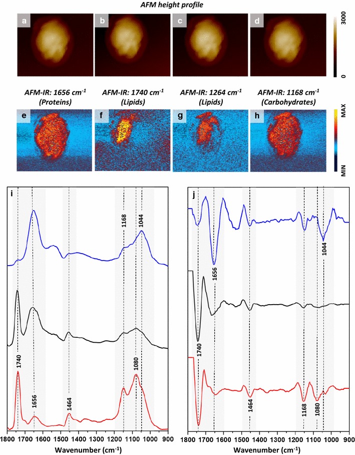
AFM–IR analysis of HBY31 cells. a–d AFM height results recorded simultaneously with e–h AFM–IR imaging of selected wavenumbers, demonstrating the distribution of chosen components: e proteins (1656 cm−1) (corresponding height: a), f lipids (1740 cm−1) (corresponding height: b), g lipids (1264 cm−1) (corresponding height: c) and h carbohydrates (cell wall, 1168 cm−1) (corresponding height: d). The size of imaged area was 5.41 × 4.47 µm. The scale bar for AFM height is given in nm. i A comparison of AFM–IR spectra and j their 2nd derivatives in the range 1800–900 cm−1 recorded from: LB (in red) of HBY31 cell, cytoplasm of HBY31 cell (in black) and cytoplasm of control cell (in blue). The spectral regions and bands showing most prominent differences were marked in grey background and black dash lines
