
Fig. 3.
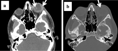
a Orbital computed tomography showing proptosis, posterior dislocation of the lens, thickening of the periorbital soft tissue and the posterior wall of the globe. b Gas bubbles in the left eye
Official websites use .gov
A
.gov website belongs to an official
government organization in the United States.
Secure .gov websites use HTTPS
A lock (
) or https:// means you've safely
connected to the .gov website. Share sensitive
information only on official, secure websites.
a Orbital computed tomography showing proptosis, posterior dislocation of the lens, thickening of the periorbital soft tissue and the posterior wall of the globe. b Gas bubbles in the left eye