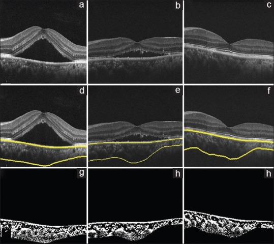
Figure 2.

Sham laser, optical coherence tomography of the right eye of a 40-year-old male using spectral domain optical coherence tomography at baseline, 3 months, and 6 months, (a-c), respectively. (d-f) Images show choroid segmentation using an automated algorithm. (g-i) are the binarized images, which is used to calculate the choroidal vascularity index (0.56, 0.58, and 0.59, respectively)
