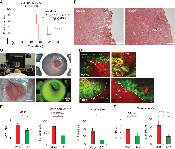
Fig. 6.
SYK modulates the tumor microenvironment in immune-competent GL261-L2G/C57BL/6J mice model. (A) Kaplan–Meier survival analysis of mock- (red) or BAY-treated (green) C57BL/6J mice orthotopically injected with the GL261-L2G murine glioma cell line; n = 7. (B) Hematoxylin and eosin staining of mock- and BAY-treated tumors. (C) Multiphoton setup, window, and epifluorescence of injected GL261-L2G cells after 7 days of implantation. (D) Multiphoton imaging of mock- or BAY-treated GL261-L2G tumors from depths 20–45 µm below the tumor surface. Lymphocyte “shadows” are marked with a white arrowhead. Bar represents 100 µm. (E) Multiphoton analysis of mock- (red) or BAY-treated (green) GL261-L2G tumors showing morphology (or invasiveness) of tumor cells (length/width ratio), number of dextran-labeled phagocytic cells, and unlabeled “shadows” (lymphocytes); n > 3 mice, n = 6–10 fields, 21 Z-slices per field (0–100 µm depth), 5 µm apart. (F) FACS analysis of infiltrated CD19+ and CD11b+ cells showing percentage of parent cells; n = 4. Error bars show SD. *P < 0.05, **P < 0.01.

