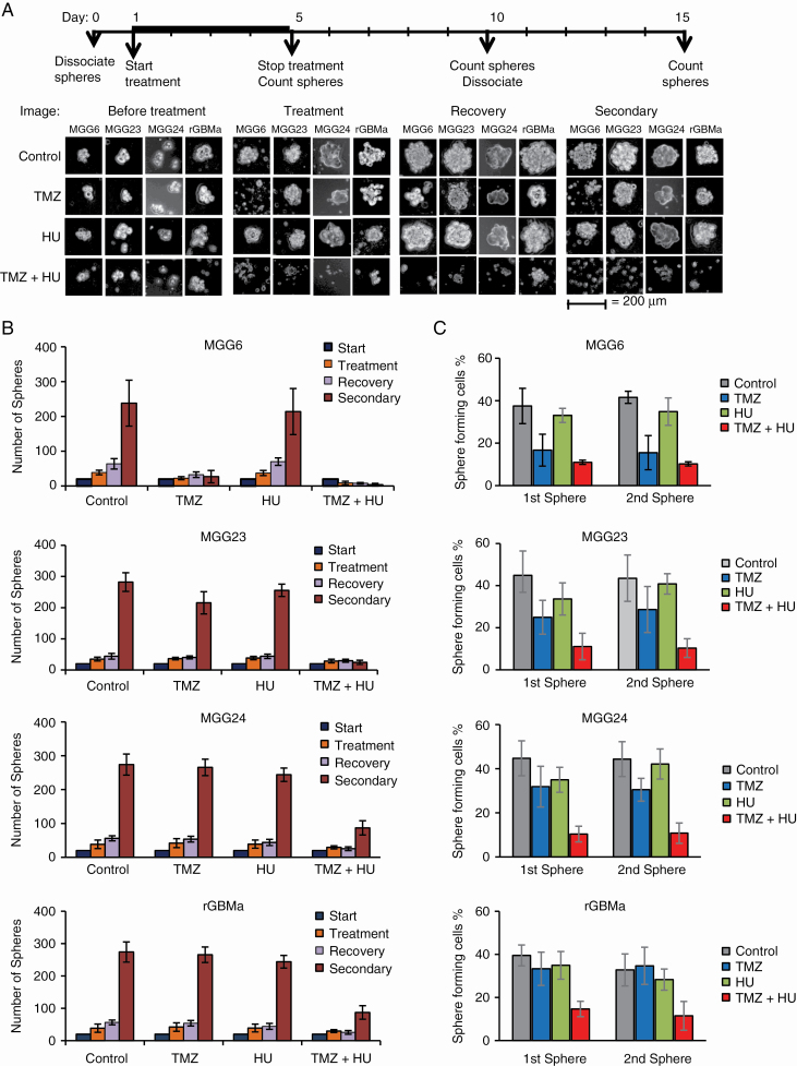
Fig. 3.
Effect of HU+TMZ on GBM neurospheres with different MGMT status, genetic background, and molecular subtype. (A–B) Neurospheres from newly diagnosed GBM tumors with methylated (MGG6), unmethylated (MGG23), or mixed (MGG24) MGMT promoter and from recurrent (rGBMa) tumors were treated with either DMSO, HU, TMZ, or HU+TMZ for 4 days. Spheres were counted and left without treatment for another 5 days to allow recovery. Recovered spheres were dissociated and 1000 cells were plated in new 48-well plates to measure secondary sphere formation 5 days later. The experiment was repeated 3 times, and a representative image from 4 replicates in each treatment group is displayed (A). Scale bar, 200 μm. Total sphere numbers in the well recorded at each event; *P < 0.05, **P < 0.01 vs TMZ alone (A). (C) Sphere limiting dilution analysis comparing stem cell frequency. Different amounts (1 to 1000 cells) of GBM stem cells were plated as single cells. The number of cells that could form a sphere were quantified at day 5 (first sphere) and day 15 (second sphere) as described in the Materials and Methods section; *P < 0.05 TMZ+HU vs TMZ.

