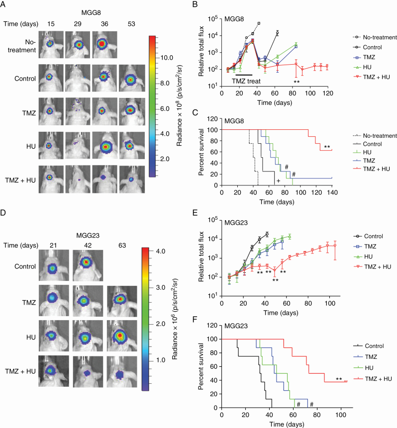
Fig. 4.
HU sensitizes patient-derived GBM tumors to TMZ in vivo irrespective of MGMT status. (A–C) 5×10⁴ MGG8 cells with methylated MGMT promoter expressing Fluc were stereotactically injected into the left mid-striatum of nude mice brain as small neurospheres (day 1). Four weeks later, mice were divided into 2 groups and treated with either TMZ (n = 34) or vehicle (n = 6) for 2 weeks, and then left off-treatment for 3 weeks. The TMZ-treated group was then divided into 4 subgroups, which received vehicle, HU, TMZ, or HU+TMZ (n = 8–10/group). Representative Fluc images of a single mouse from each group at different time points are shown (A). Tumor-associated Fluc signal was quantified (B); **P < 0.01, TMZ+HU vs TMZ only. Animal survival was recorded in a Kaplan–Meier plot (C); +P < 0.05 control vs no-treatment, #P < 0.05 vs control, **P < 0.01 TMZ+HU vs TMZ only by log-rank test. (D–F) MGG23 cells with unmethylated MGMT promoter were implanted in the brain of nude mice and treated with DMSO, HU, TMZ, or TMZ+HU. (D) Representative Fluc images of a single mouse from each group are shown over time. (E) Quantitation of tumor-associated Fluc signal; **P < 0.01 TMZ+HU vs TMZ. (F) Kaplan–Meier plots analyzing survival, #P < 0.05 vs control, **P < 0.01 TMZ+HU vs TMZ only.

