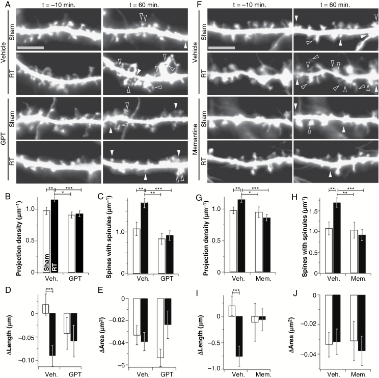
Fig. 2.
GPT and memantine prevent acute radiation-induced changes in live dendrite morphology. (A) Representative images of segments of secondary dendrites from vehicle- and GPT (5 units/mL)-treated neurons at the indicated time points after both sham treatment and irradiation (RT). Filled arrowheads indicate newly formed spines and open arrowheads indicate newly formed spinules and extra spine heads. GPT prevented increases in projection density (B) and spinules (C), as well as decreases in spine length (D). Data are shown as means ± SEM (n = 261–452 spines/13 neurons, N = 3). *P ≤ 0.05, **P ≤ 0.01, ***P ≤ 0.001. Bar is 5 µm. (F–J) The same experiment as in panels A–E was repeated using 25 µM memantine in place of GPT (n = 221–461 spines/13 neurons, N = 3). *P ≤ 0.05, **P ≤ 0.01, ***P ≤ 0.001. Bar is 5 µm.

