
Figure 1.
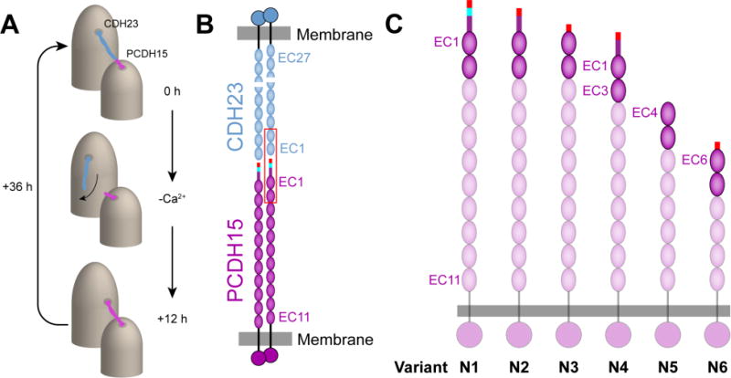
Model of the tip link interaction and structural details of the splice variants of PCDH15. (A) The tip link is composed of CDH23 and PCDH15 and connects adjacent stereocilia located on the apical side of hair cells. Upon removal of calcium, the tip link is disrupted and forms short-lived PCDH15-PCDH15 links.12 (B) Each homodimer of CDH23 and PCDH15 bind together to form an antiparallel heterotetrameric complex. The first two EC repeats located at the N-terminus of each protein form a mechanically strong bond (outlined in red). (C) PCDH15 is present in at least 26 alternative splice variants, and a subset of these variants has alterations in the first two EC repeats (opaque ovals). These variants have been grouped into 6 categories numbered N1-N6 (Figure S1). The start of the N-terminus is drawn as a rectangle where the end of exon 2 is indicated in red, exon 3 is shown in cyan, and exon 4 is shown in purple.