
Figure 3.
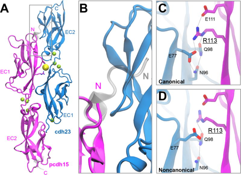
Structure of a noncanonical cdh23-pcdh15 complex. (A) X-ray crystal structure of cdh23 and pcdh15(N2) (PDB: 4XXW) overlaid with the canonical cdh23 and pcdh15(N1) (PDB: 4AQ8) complex (shown in gray). (B) Region of gray box in panel (A), highlighting important structural differences at the N-terminus of pcdh15. (C) Detail of the interface between cdh23 and pcdh15 shows residue R113 pointing towards the interface in the canonical structure. (D) In the noncanonical structure, R113 is oriented away from the interface and forms fewer favorable interactions with nearby residues.