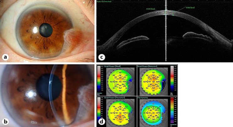
Fig. 3.
Clinical images of the patient on day 26. a Slit-lamp examination showed that cell infiltration had disappeared. b Slight opacification was left at the center of cornea. c AS-OCT showed that the wound had closed and corneal edema had improved. d AS-OCT analysis suggested that the corneal irregularity caused by the wound was small.

