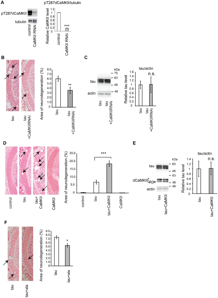
Fig. 1.
Knockdown of CaMKII suppresses, while overexpression of CaMKII promotes, neurodegeneration induced by tau. (A) CaMKII RNAi reduces active CaMKII in Drosophila brain. Western blot analysis of fly heads carrying the pan-neuronal elav-Gal4 driver alone (control) or expressing CaMKII RNAi driven by elav-Gal4 (CaMKII RNAi) with anti-pT286 CaMKII antibody. Tubulin was used as a loading control. Mean ± SD, n = 5, ***, P < 0.005, Student’s t-test. (B) RNAi-mediated knockdown of CaMKII suppresses tau-induced neurodegeneration. (Left) The lamina of flies expressing human tau alone (tau) and co-expressing human tau and CaMKII RNAi (tau+CaMKIIRNAi) driven by GMR-Gal4. Neurodegeneration is indicated by arrows. (Right) Quantification of neurodegeneration, Mean ± SEM, n = 8–12. **, P < 0.01. (C) RNAi-mediated knockdown of CaMKII does not change tau protein levels. Western blot analysis of fly heads expressing human tau alone (tau) and co-expressing human tau and CaMKII RNAi (tau+CaMKIIRNAi) driven by GMR-Gal4 with anti-tau antibody. Actin was used as a loading control. Mean ± SD, n = 5, n.s., P > 0.05. (D) Overexpression of CaMKII enhances tau-induced neurodegeneration. (Left) The lamina of control flies bearing the GMR-Gal4 driver only (control), flies expressing tau alone (tau), co-expressing tau and CaMKII (tau+CaMKII) and CaMKII alone (CaMKII). (Right) Quantification of neurodegeneration. Mean ± SEM, n = 8–12. ***, P < 0.005. (E) Overexpression of CaMKII does not change tau protein levels. Western blot analysis of fly heads expressing human tau alone (tau) and co-expressing human tau and CaMKII (tau+CaMKII) driven by GMR-Gal4 with anti-tau antibody (tau). Expression of exogenous CaMKII (R3) is confirmed by Western blot with anti-dCaMKII antibody (dCaMKII). Please note that this blot reflects all the CaMKII proteins in the head including endogenous CaMKII. Endogenous CaMKII is expressed in multiple isoforms and abundant in all the brain regions, while exogenous CaMKII is R3 isoform (arrowhead) and expressed only in the retina. Mean ± SD, n = 5, n.s., P > 0.05. (F) Inhibition of CaMKII activity suppresses tau-induced neurodegeneration. (Left) The lamina of flies expressing human tau alone (tau) and co-expressing human tau and the inhibitory domain of the rat CaMKII (tau+ala) driven by GMR-Gal4. (Right) Quantification of neurodegeneration. Mean ± SEM, n = 8–12. *, P < 0.05.

