
Fig. 1.
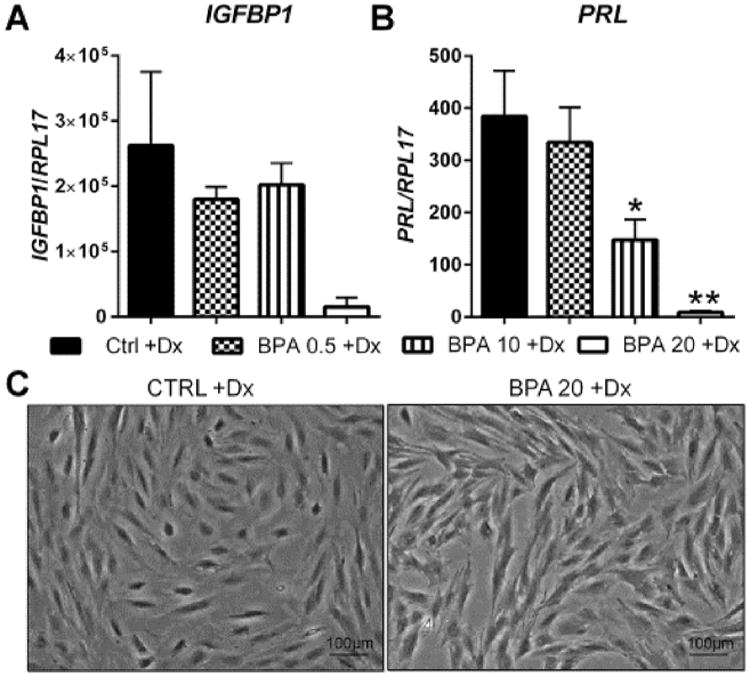
Effect of BPA on in vitro decidualization of uterine stromal fibroblasts. HuF cells were exposed to BPA at several doses (0.5 μg/mL, 10 μg/mL and 20 μg/mL) in the presence of a decidualization-inducing hormonal formulation for eight days. At the conclusion of the treatment, IGFBP1 (A) and prolactin (B) mRNA expression was determined by qRT-PCR which was normalized to the expression of RPL17. Morphological changes (C) were observed in HuF cells treated with 20 μg/mL of BPA as compared to cells with hormone treatment alone. Data are presented as mean ± SEM from four independent experiments from four different cell lines. (*p <0.05, **p <0.01 compared to decidualized controls). Note: Dx in Fig. stands for decidualization.