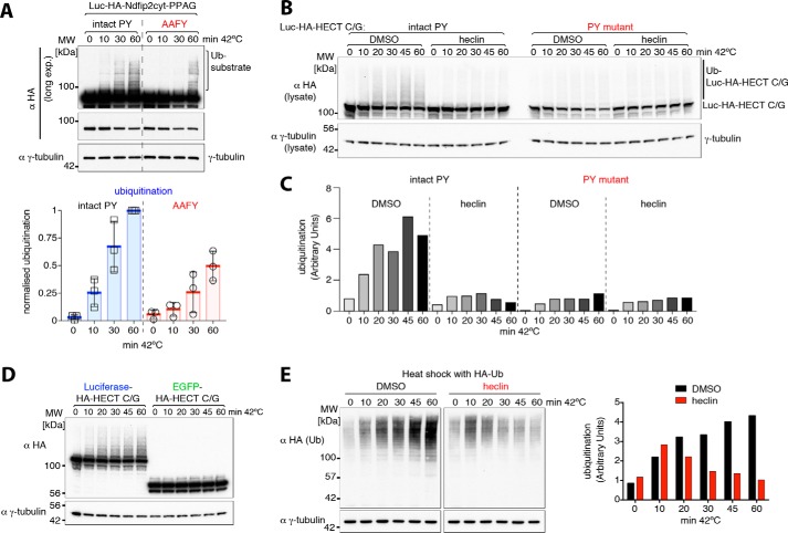
Figure 5.
Role of heat shock–exposed “inaccessible” PY motifs in ubiquitination. A, Western blots of whole-cell lysates from control or heat-shocked HEK293T cells co-transfected with His-Ub and luciferase-Ndfip2 (3xPPAG)-EGFP or a mutant luciferase version (AAFY). Quantification of ubiquitinated luciferase constructs via densitometry is shown on the right, showing reduced and delayed ubiquitination of the AAFY mutant upon heat shock. MW, molecular weight. B, Western blots of whole cell lysates from control or heat-shocked HEK293T cells co-transfected with His-Ub and luciferase-fused inactive HECT domain with an intact or mutant (normally inaccessible) PY motif. Heat shock experiments were carried out either with 1% DMSO control or 100 μm heclin as indicated. C, quantification of substrate ubiquitination via densitometry shown in B. D, Western blots of whole-cell lysates from control or heat-shocked HEK293T cells co-transfected with His-Ub and either luciferase or EGFP-fused inactive HECT domain, showing luciferase-dependent ubiquitination upon heat shock. E, HEK293T cells expressing hemagglutinin (HA)-Ub were treated with DMSO control or heclin and harvested after incubation at 42 °C for the times indicated. A densitometric quantification of ubiquitination is shown on the right. All blots are representative of two or three similar independent experiments, and the graph in A shows individual data points from different experiments, with the mean and standard deviation indicated. Densitometric quantification data were obtained within a linear range of exposure, and ubiquitination levels were normalized to the highest value in each set of experiments.

