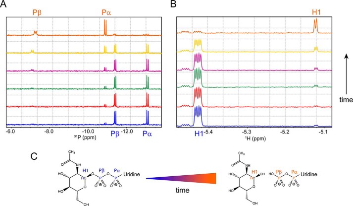
Figure 4.
SseK3(14–335) hydrolyzes UDP-GlcNAc in UDP and free N-acetyl α-d-glucosamine. A and B, phosphorus (A) and proton (B) NMR spectra of a 500 μm solution of UDP-GlcNAc in the presence of 10 μm SseK3(14–335). The spectra were recorded at different time points: 0 min (blue), 5 min (red), 15 min (green), 30 min (purple), 90 min (yellow), and 12 h (orange). C, UDP-GlcNAc hydrolysis reaction. The Pα and Pβ doublets and the H1 multiplets are assigned in the relative spectra and highlighted on the chemical structures of the ligand in its intact and hydrolyzed form.

