Figure 1.

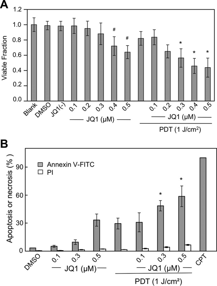
Cytotoxic effects of PDT on glioblastoma U87 cells: Enhancement by BET bromodomain inhibitor JQ1. A, cells at ∼60% confluency were either treated directly with JQ1 in increasing concentrations up to 0.5 μm or treated with JQ1 after preincubation with 1 mm ALA followed by irradiation (light fluence ∼ 1 J/cm2). Nontreated (blank), and vehicle DMSO- and 0.5 μm JQ1(−)-treated cells were studied alongside as controls. After 24 h of dark incubation, cell viability was determined by CCK-8 assay. Plotted values are means ± S.E. (n = 4); *, p < 0.05 versus PDT alone or 0.3 μm JQ1 alone; #, p < 0.05 versus blank or DMSO vehicle control. B, cells prepared as described in A were analyzed for extent of apoptosis or necrosis, 5 h after treatment with JQ1 or PDT plus JQ1, using annexin V–FITC for apoptosis and propidium iodide for necrosis. Camptothecin (CPT, 25 μm) served as an indicator for maximum apoptosis. Plotted data are means ± S.E. (n = 4); *, p < 0.01 versus PDT alone or 0.3 μm JQ1 alone.
