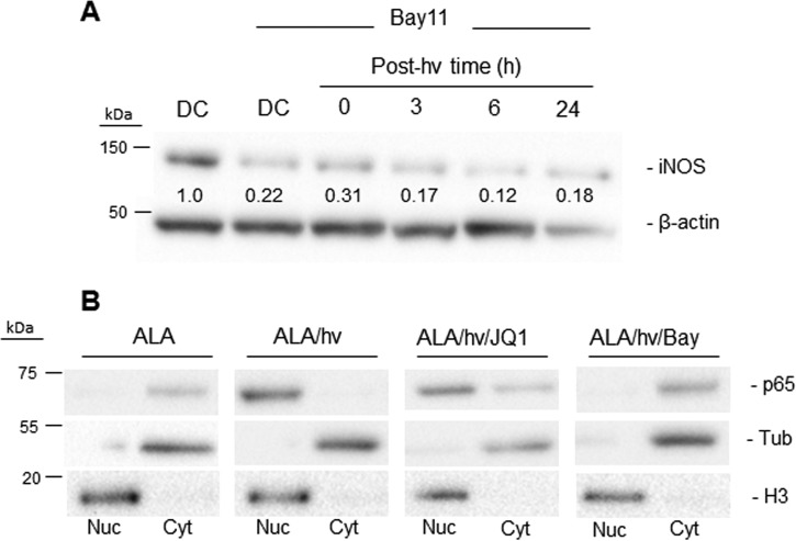
Figure 4.
NF-κB involvement in post-PDT up-regulation of iNOS in glioblastoma cells. A, U87 cells were sensitized with PpIX by preincubation with 1 mm ALA for 30 min. After washing, the cells were treated with Bay11 and either dark-incubated for 24 h (DC) or irradiated (1 J/cm2) and then analyzed for iNOS and β-actin by immunoblotting after increasing post-irradiation times up to 24 h. A dark control without Bay11 was also analyzed. Numbers below the bands are iNOS/β-actin ratios relative to DC (minus Bay11). hν, light. B, NF-κB activation and subcellular distribution following PDT. U87 cells treated with PDT alone (ALA/hν), PDT plus 0.3 μm JQ1 (ALA/hν/JQ1), or PDT plus 5 μm Bay11 (ALA/hν/Bay) were homogenized at 5 h after irradiation and separated into nuclear (Nuc) and cytosolic (Cyt) fractions, each of which was immunoblotted for the p65 subunit of NF-κB along with histone H3 as a nuclear marker and α-tubulin (Tub) as a cytosolic marker. A dark control (ALA-only) was analyzed similarly.

