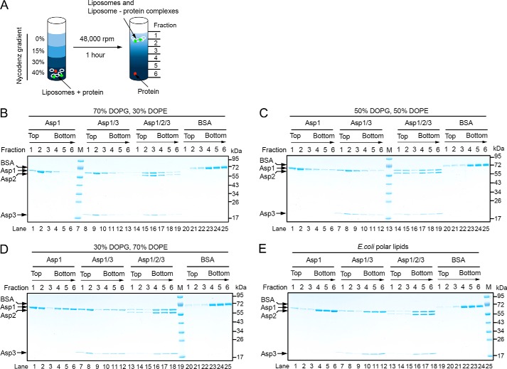
Figure 6.
Interaction of the Asps with phospholipid bilayers. A, scheme of the binding assay. Liposomes containing different lipid compositions were mixed with Asps, and the samples were subjected to flotation in a Nycodenz gradient. Fractions were collected from the top and analyzed by Coomassie staining. Liposome-bound proteins (green) are expected to co-float with the lipids (white). Non-associated proteins (red) stay at the bottom. B–D, indicated combinations of Asps were tested for binding to liposomes containing a different ratio of the negatively charged lipid DOPG and the neutral lipid DOPE. Lipids peak in fraction 2. Bovine serum albumin (BSA) was used as a control. E, binding of the Asps to liposomes generated with E. coli polar lipids.

