. 2018 Jan 15;32(1):101–108. doi: 10.21873/invivo.11210

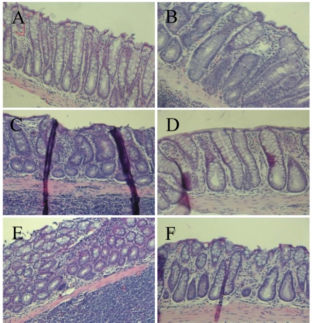
Figure 2. Histopathological examination of colonic tissues of F344 rats from group 1, saline + control diet (A); group 2, azoxymethane (AOM) + control diet (B); group 3, AOM + 1% Capsosiphon fulvescens-supplemented diet (C); group 4, AOM + 2% C. fulvescens-supplemented diet (D); group 5, AOM + 2% Hizikia fusiforme-supplemented diet (E); and group 6, AOM + 6% H. fusiforme-supplemented diet (F). Representative microscopic examination was performed with hematoxylin and eosin staining. Histological assessment showed the presence of aberrant crypt foci including dysplasia and hyperplasia, abnormally-shaped lumens and elongated nuclei in the colonic mucosa of the treated control group but a marked reduction in all experimental groups. Sections were examined at 200× magnification.

Figure 2