Figure 3.
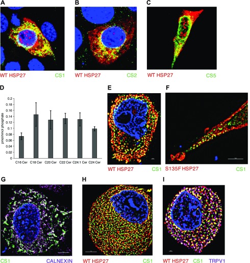
Hsp27:CerS1 interaction localizes to the ER. A–C) WT Hsp27 (red) colocalizes with FLAG-tagged CerS1, CerS2, and CerS5, respectively, (green) in HT-22 cells by confocal microscopy. D) Ceramide profile from DRGs cultured for 4 d from 7 mo female mice (n = 3). E, F) WT and S135F Hsp27 (red), respectively, colocalize with FLAG-tagged CerS1 (green) in HT-22 cells by SIM microscopy. G) FLAG-tagged CerS1 (green) and calnexin (pink) strongly colocalize. H) Endogenous WT-Hsp27 (red) and anti-CerS1 (green) colocalize in mouse DRG. I) Electroporated S135F Hsp27 (red) and FLAG-tagged CerS1 (green) colocalize in mouse DRG cells. Confirmation of neuronal identity with TrpV1 (pink).

