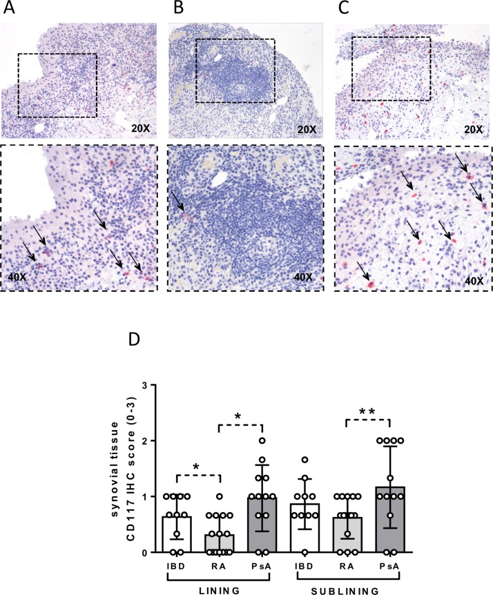
Figure 3.
Immunohistochemical (IHC) staining for CD117 on synovial tissue (ST) of patients with inflammatory bowel disease (IBD) with onset of peripheral arthritis under tumour necrosis factor inhibitor (TNF-i), patients with rheumatoid arthritis (RA) naive to treatment and patients with psoriatic arthritis (PsA) naive to treatment. (A) CD117 (red) staining of ST from patient with IBD with onset of peripheral arthritis under TNF-i treatment (magnification 20× and 40× for the corresponding insert) (thin black arrows indicate CD117+ cells). (B) CD117 (red) staining of ST from patient with RA naive to treatment (magnification 20× and 40× for the corresponding insert) (thin black arrow indicates CD117+ cells). (C) CD117 (red) staining of ST from patient with PsA naive to treatment (magnification 20× and 40× for the corresponding insert) (thin black arrows indicate CD117+ cells). (D) Lining and sublining IHC score for CD117+ cells in patients with IBD (n=10) with onset of peripheral arthritis under TNF-i, patients with RA naive to treatment (n=15) and patients with PsA naive to treatment (n=12); lining IHC CD117 score in patients with IBD versus patients with RA naive to treatment, *p<0.02; lining IHC CD117 score in patients with RA naive to treatment versus patients with PsA naive to treatment, *p<0.02; sublining IHC CD117 score in patients with RA naive to treatment versus patients with PsA naive to treatment, **p<0.05.

