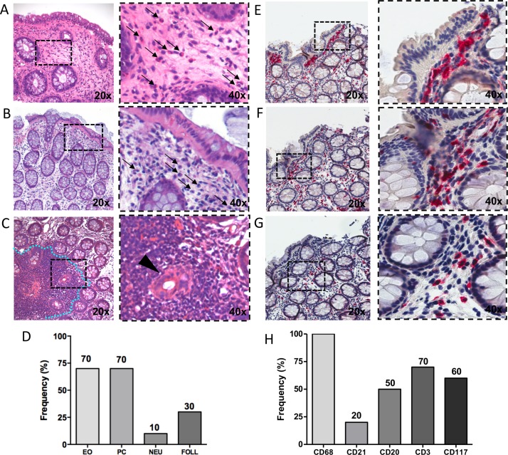
Figure 4.
H&E and immunohistochemical (IHC) staining for CD68/CD21, CD3/CD20 and CD117 on colonic mucosa of patients with inflammatory bowel disease (IBD) in disease remission/low disease activity with onset of peripheral arthritis under TNF-i. (A) H&E staining of colonic mucosa of patient with IBD in disease remission with onset of peripheral arthritis under TNF-i showing the presence of eosinophils (thin black arrows). (B) Plasma cells (thin black arrows) and (C) gut follicles (blue dotted line) and cripts damage (back arrow head) (magnification 20× and 40× for the corresponding insert). (D) Frequency of eosinophils (EO), plasma cells (PC), neutrophils (NEU) and gut follicles (FOLL) presence in the colonic mucosa biopsies of patients with IBD (n=10) in disease remission/low disease activity with onset of peripheral arthritis under TNF-i. (E) CD68 (red)/CD21 (brown) staining of colonic mucosa from patient with IBD with onset of peripheral arthritis under TNF-i treatment. (F) CD3 (red)/CD20 (brown) staining of colonic mucosa from patient with IBD with onset of peripheral arthritis under TNF-i treatment. (G) CD117 (red) staining of colonic mucosa biopsy from patient with IBD with onset of peripheral arthritis under TNF-i treatment (magnification 20× and 40× for the corresponding insert). (H) Frequency of CD68+, CD21+, CD3+, CD20+ and CD117+ cells presence in the colonic mucosa biopsies of patients with IBD (n=10) in disease remission/low disease activity with onset of peripheral arthritis under TNF-i.

