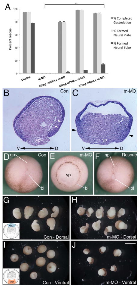
Figure 2. mRNA rescue and MO effects on convergent extension.
A) Partial rescue of m-MO phenotype by introduction of Mov10 mRNA. Error bars represent SEM, **p<0.01 (Student’s t-test, two-tailed). Mov10 mRNA rescued cases that underwent gastrulation also formed a neural plate, but those cases typically disintegrate a few hours later so they are unable to complete neurulation and form a neural tube. B–C) X. laevis stage 10 sagittal sections stained with Hematoxylin and Eosin. Dorsal (D) and ventral (V) sides are as noted. B) Representative section of a control (Con) embryo, showing Brachet’s cleft (white arrowheads), which separates the outer ectoderm from the underlying mesendoderm. C) Representative section of a sibling m-MO injected embryo, which lacks a visible delineation between ectodermal and mesendodermal layers (Brachet’s cleft). Note that cells along the dorsal and ventral sides of the blastopore and yolk plug, denoted with black arrowheads, appear to be thicker, having mainly undergone convergent thickening (compare B to C). D) Whole-mount image of representative stage 13–14 Control (Con) embryo with closed blastopore. E) Representative sibling m-MO embryo with unclosed blastopore. F) Rescued embryo co-injected with m-MO and Mov10 mRNA (250pg) with closed blastopore. G–J). Development of stage 10 dorsal (dmz) and ventral marginal zone (vmz) explants harvested from the region shown in the inset included in G and I, respectively. G) Control dmz explants eventually form mesoderm that undergoes convergent extension, as revealed by the formation of elongated projections. H) dmz explants from m-MO injected embryos also show signs of convergent extension and elongation. Note, however, that the pigmented ectoderm does not cover the mesodermal tissue as completely as seen in control explants (compare with G). I) vmz explants harvested from stage 10 control embryos do not exhibit signs of elongation and form spherical embryoids. J) Likewise, m-MO injected embryos also form spherical embryoids. Note that the pigmented ectoderm has not covered the surface of these explants. bl, lip of blastopore; np, neural plate; yp, yolk plug. Scale bar in J equals 180μm for B–C, 250μm for D–F, 850μm for G–J.

