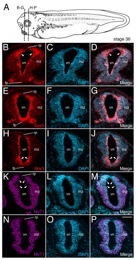
Figure 7. Knockout of zygotic Mov10 shows abnormal staining of neuronal precursors in the ventricular zone.
A) A stage 36 tadpole showing the plane of sectioning for B through P. B) Representative fluorescence image of control embryo shows the forebrain region with Sox3 positive precursors (in red) surrounding the lumen of the ventricle. Note the expanded dorsal Sox3 labeling in the ventricular zone, denoted by white arrowheads. C) DAPI staining (in blue) of the same section shown in B. D) Merged images from B and C. E–G) Representative fluorescence images from a similar region, as shown in B, from representative z-MO injected tadpoles. E) Sox3 positive neuronal precursor cells are located in the ventricular zone. F) DAPI staining of the same section shown in E. G) Merged images from E and F. Notice the enhanced overall staining of Sox3, including in the more ventral regions of the ventricular zone, denoted by white arrowheads (compare to the control embryo shown in B). H–J) More posterior section from the forebrain region of a z-MO injected tadpole. H) Sox3 neuronal precursors. Note the expanded ventral labeling denoted by arrowheads. I) Corresponding DAPI staining for H. J) Merge of Sox3 and DAPI stains. K–M) MyT1 staining for differentiated neurons in a control embryo. K) MyT1 positive differentiated neurons. Note expanded dorsal area devoid of MyT1 expression within the ventricular zone (denoted by white arrowheads). L) DAPI staining of the same section shown in K. M) Merged images from K and L. N–P) Representative sections from a z-MO injected tadpoles. N) Wide distribution of MyT1 positive differentiated neurons. Note uniform MyT1 staining in the dorsal ventricular zone (compare with K). O) DAPI staining of the same section shown in N. P) Merged images from N and O. fp, floor plate; mz, marginal zone; rp, roof plate; vn, ventricle. Scale bar in P equals 40μm

