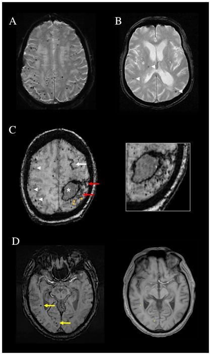
Figure 1.
Patterns of cerebral microbleeds (CMB). (A) Multiple strictly lobar CMB on T2*-weighted MRI of a 69-year-old woman who presented with a spontaneous lobar intracerebral hemorrhage. Brain autopsy showed advanced CAA. (B) Mixed CMB (arrowheads) affecting the right thalamus, a deep hemispheric territory, as well as lobar brain regions and therefore not fulfilling Boston criteria for probable CAA. (C) Subacute left frontoparietal lobar hemorrhage (asterisk), numerous strictly lobar CMB (white arrowheads), and a left frontal focus of cortical superficial siderosis (white arrow) on susceptibility-weighted imaging (SWI) MRI of a 78-year-old woman. The image additionally shows foci of CMB (orange arrowheads) and cortical superficial siderosis (red arrows, also seen in magnified image) that are immediately adjacent to the lobar hemorrhage and therefore not counted as separate lesions in determining the number of lobar hemorrhagic foci. (D) SWI from a 71-year-old man with memory loss and CAA on brain biopsy. The yellow arrows point to CMB in the right temporal and right occipital lobes that might appear distant from the brain surface, but would be counted as lobar microbleeds. The aligned T1-weighted slice shows that their positions (asterisks on the right panel) are within or very close to the cortical ribbon.

