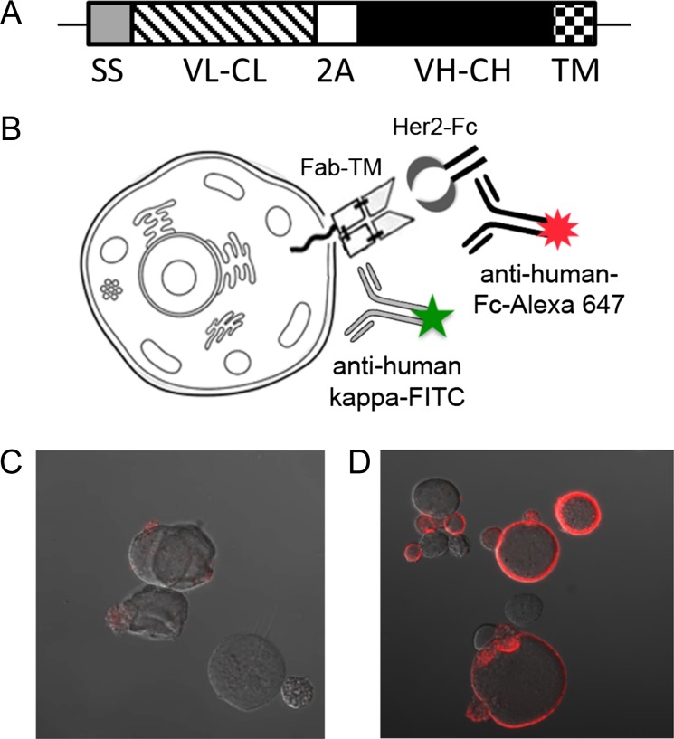
Fig. 1.
The hu4D5 Fab was effectively displayed on the surface of CHO cells. (A) Schematic of the Fab CHO expression construct. The open reading frame inserted into pPyEBV consisted of a murine IgΚ secretion signal (SS) fused to the hu4D5 IgΚ chain (VL–CL) DNA sequence, followed by DNA encoding a furin cleavage site and F2A peptide (2 A). The hu4D5 VH and CH1 coding sequence (VH–CH) fused to a short glycine–serine linker and the PDGFR transmembrane domain (TM) immediately followed the 2A peptide. (B) Schematic of the Fab CHO display and staining system. The displayed Fab was stained with HER2-Fc, then anti-human Fc-Alexa Fluor 647, and anti-human IgK-FITC was added for flow cytometric detection. For microscopy visual confirmation of the Fab CHO expression and staining procedure, CHO cells were transfected with either (C) blank pPyEBV or (D) pPyhu4D5disp and stained for HER2 binding, then imaged by confocal microscopy.

