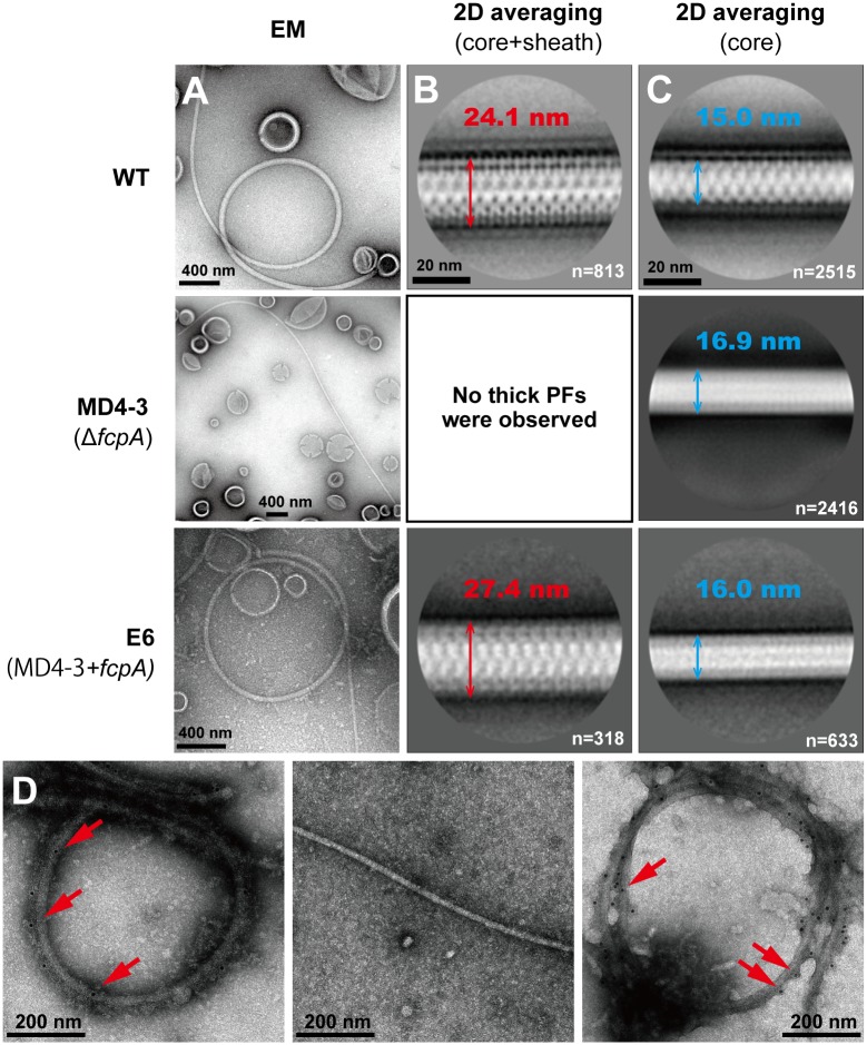
Fig 1. Morphological and immunological characterizations of purified PFs from the wild-type (WT), slow-motility mutant MD4-3 (ΔfcpA), and complemented (E6) strains of L. biflexa.
A. Transmission electron microscopic images of negatively stained, purified PFs. B. The diameters of averaged thick PF (core + sheath) images. n: number of particles used for averaging. C. The diameters of averaged thin PF (core) images. n: number of particles used for averaging. D. Immunoelectron microscopic images of purified PFs labeled with anti-FcpA antiserum. Left: WT, middle: MD4-3, right: E6. Arrows indicate 10 nm gold nanoparticles conjugated to secondary antibody.

