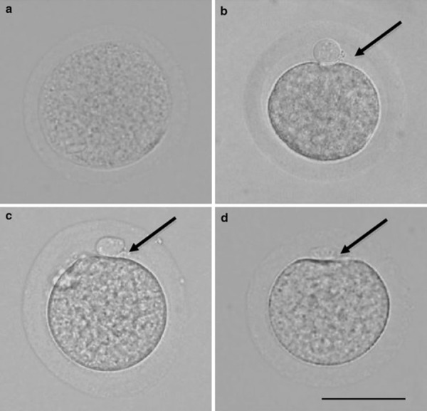
Figure 4.

Hamster oocytes. Scale bar shows 60 μm. Arrows indicate the perivitelline space. a Oocyte immediately after collection from antral follicle. b Oocyte immediately after collection from oviduct. c Oocyte cultured for 16 h in mTALP3 medium. d Oocyte cultured for 16 h in mTALP3 medium with 0.25 mm 4‐methylumbelliferone
