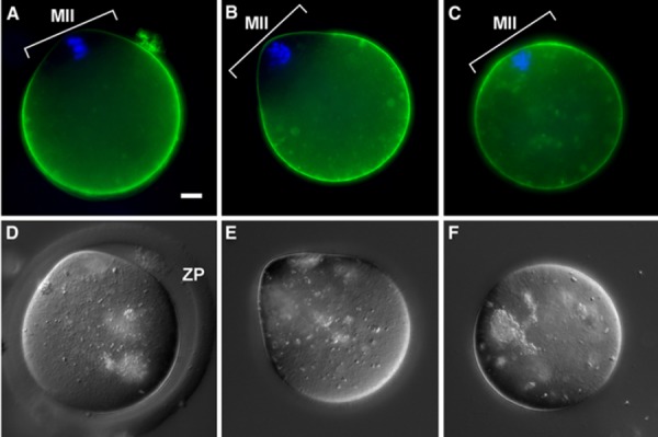
Figure 2.

Effect of collagenase and acidified Tyrode's solution treatment on egg membrane molecule distribution observed by CD9‐EGFP localization. a–c Fluorescent image with CD9‐EGFP transgenic mouse eggs to trace the damage of oolemma. Green CD9‐EGFP. Blue Hoechst 33342 staining for chromosomes. d–f DIC images. a, d Control (ZP intact without cumulus cells after hyaluronidase treatment but before ZP removal treatment). b, e Collagenase treatment; one‐step collagenase method. c, f Acidified Tyrode's solution treatment. In the control (a) and collagenase treatment (b), CD9 is not localized (normal) on the oolemma overlying the MII chromosome area (MII), whereas in acidified Tyrode's solution treatment (c), CD9 was diffusely distributed even on the oolemma overlying the MII chromosome area; no polarization of CD9 was found; however, polarity was apparent in the control (a) and collagenase treatment (b). ZP Zona pellucida. Bar 10 μm
