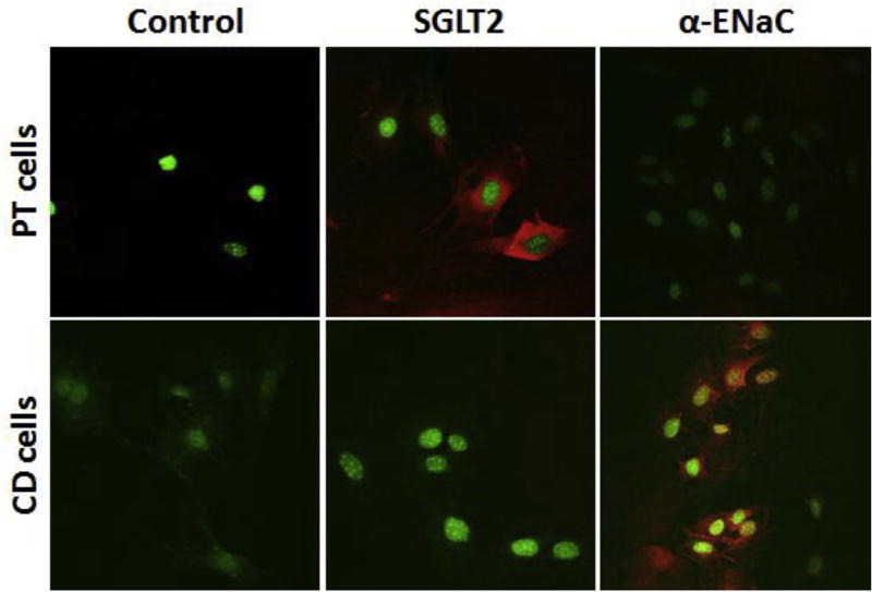
Fig. 2.
Immunofluorescence staining showing the expression of marker protein for proximal tubule or collecting duct cells: Mouse proximal tubule (PT) cells or collecting duct (CD) cells were freshly isolated from the mouse kidney via the Miltenyi Biotec's Magnetic-Activated Cell Sorting (MACS) technology. We used biotinylated Lotus tetragonolobus lectin antibody (Vector #B-1325) for the separation of PT cells and Dolichos biflorus agglutinin antibody (Vector #B-1035) for separation of CD cells. The purity of the cells prepared was assessed by immunofluorescence staining using the antibody to the SGLT2 (a marker protein of PT cells, S1 and S2 segments) and the antibody to α-ENaC (marker protein of CD cells). Red stain indicates the presence of SGLT2 or α-ENaC in PT or CD cells respectively. DAPI-stained nuclei are shown in green.

